Reovirus Picture Gallery
Jump To: Mammalian Reovirus Virion Surface Proteins | Mammalian Reovirus Core Surface Proteins
More Mu1 and Sigma3 | Viral Factories
-
Back to Top
Mammalian Reovirus Virion Surface Proteins
-
Click thumbnail to view larger image.
Rendered with Rasmol 2.7.1. Peptide chains: cartoon format, alpha helices in hot pink, beta sheets in yellow. Heteroatoms: spacefilling format, zinc ions in black, beta-octylglucoside molecules in violet. Structures from Reinisch et al. (2000) (1EJ6.pdb), Liemann et al. (2002) (1JMU.pdb), and Chappell et al. (2002) (1KKE.pdb).
Back to Top
Mammalian Reovirus Core Surface Proteins
-
Click thumbnail to view larger image.
Rendered with Rasmol 2.7.1. Peptide chains: cartoon format, alpha helices in blue, beta sheets in sea green. Heteroatoms: spacefilling format, zinc ions in black. Structures from Reinisch et al. (2000) (1EJ6.pdb).
Back to Top
More Mu1 and Sigma3
-
Click thumbnail to view larger image.
Rendered with Rasmol 2.7.1. Peptide chains: cartoon format, alpha helices in hot pink, beta sheets in yellow. Heteroatoms: spacefilling format, zinc ions in black, beta-octylglucoside molecules in violet. Structures from Olland et al. (2001) (1FN9.pdb) and Liemann et al. (2002) (1JMU.pdb).
Back to Top
Viral Factories
-
Click thumbnail to view larger image.
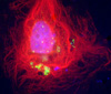
T1L |
T1L reovirus infection in CV-1 cells, stained with anti-µ2 (red) and anti-ubiquitin (green). Nuclei are stained with DAPI (blue).
|
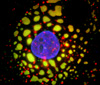
T3D
|
T3D reovirus infection in CV-1 cells, stained with anti-µ2 (green) and anti-ubiquitin (red). Nuclei are stained with DAPI (blue). |
Back to Top
|
|


